Why MRIs are invaluable to Chiropractors
We chiropractors are known for a hands-on approach to diagnosing and treating musculoskeletal issues, particularly those related to back pain and neck pain. While traditional methods such as physical exams and X-rays have long been staples in chiropractic care, the use of Magnetic Resonance Imaging (MRI) has become increasingly prevalent. This imaging technology provides a comprehensive view of the spine and surrounding tissues, enhancing our ability to diagnose and treat conditions more effectively.
At both Coventry Back Pain Clinic and Central Chiropractic Clinic we have a number of MRI providers we recommend to our patients. Each one provides exceptional reporting, clear imagining and a rapid service – on the NHS it is common to wait many months.
Understanding the Spine
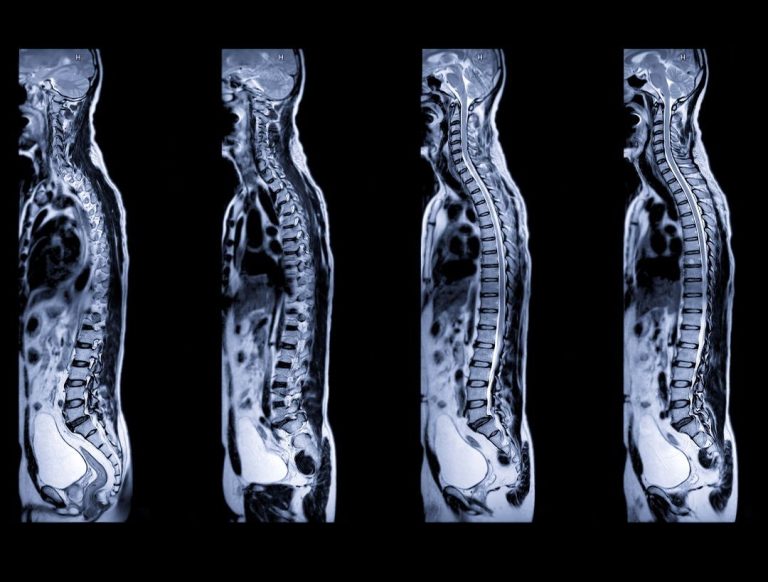
The human spine is a complex structure composed of 33 vertebrae, intervertebral discs, ligaments, and nerves. It is divided into several regions: cervical (neck), thoracic (upper and mid-back), lumbar (lower back), sacral, and coccygeal (tailbone). Each region can be a source of pain and discomfort, commonly referred to as ‘back pain’ or ‘neck pain’. The intricate nature of the spine makes accurate diagnosis crucial for effective treatment.
The role of MRIs in Chiropractic Care
1. Detailed Visualisation: Unlike X-rays, which primarily show bones, MRIs provide detailed images of both bones and soft tissues, including discs, muscles, and nerves. This comprehensive view is essential for diagnosing issues such as herniated discs, spinal stenosis, and nerve impingement, which may not be visible on standard X-rays.
2. Accurate Diagnosis: For patients suffering chronic back pain or neck pain, an MRI can reveal the underlying cause, such as a slipped disc or spinal degeneration. Accurate diagnosis is the first step towards effective treatment, and MRIs offer unparalleled detail and clarity. This is why we recommend MRIs at clinics such as Heath Lodge in Solihull, for patients experiencing long term conditions.
3. Monitoring Progress: For ongoing conditions, MRIs allow chiropractors to monitor the progress of treatment. By comparing images over time, we can assess whether the spine is healing and adjust treatment plans accordingly.
4. Avoiding Invasive Procedures: In some cases, the detailed information provided by an MRI can help chiropractors develop non-invasive treatment plans, potentially avoiding the need for surgery. For instance, understanding the precise nature of a
herniated disc can lead to targeted spinal adjustments and physical therapy, alleviating pain without surgical intervention. Only this week at our Coventry clinic on Park Road, such a scenario unfolded. A lady in her 50s was due for spinal surgery but with a second opinion via MRI we were able to establish that surgery was not required.
Common conditions identified by MRIs
- Herniated Discs: These occur when the soft centre of a spinal disc pushes through a crack in the tougher exterior casing. This can cause significant back pain and neck pain, often requiring precise imaging to identify.
- Spinal Stenosis: This is a condition involving the narrowing of spaces within the spine, which can put pressure on the nerves. MRIs are invaluable in determining the extent and location of the narrowing.
- Nerve Impingement: When nerves are compressed or pinched by vertebrae or discs, it can lead to pain, numbness, and weakness. MRIs help pinpoint the exact location of the impingement.
What to expect from the MRI scan itself?
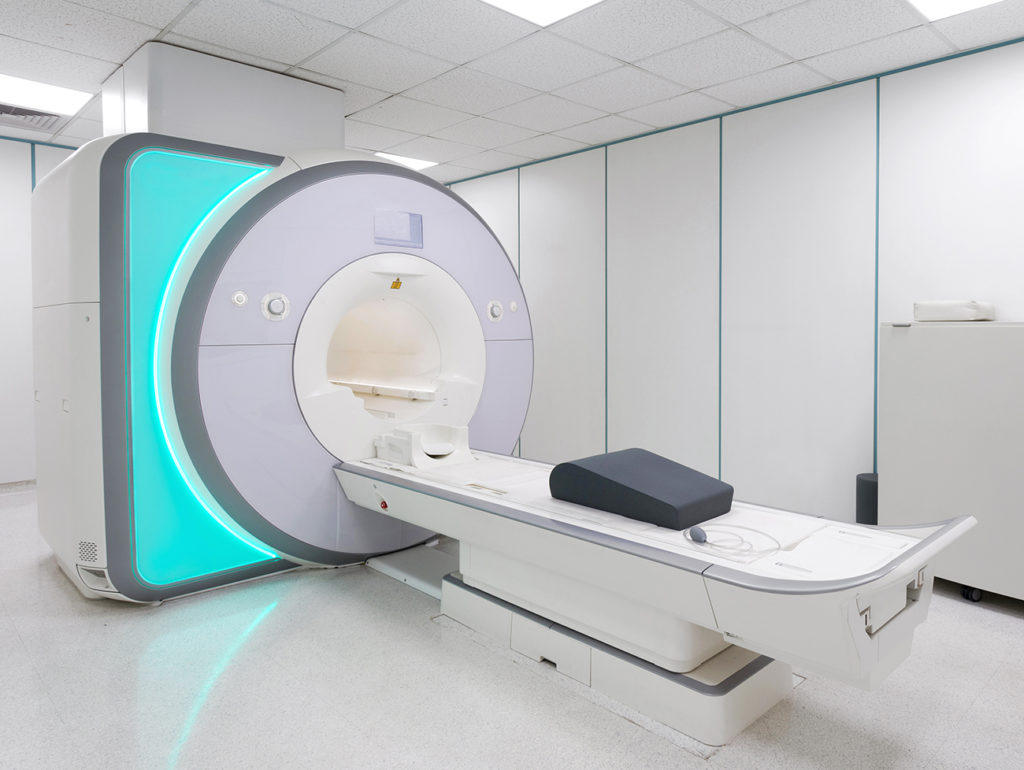
Quite often, patients who have not experienced an MRI scan will be apprehensive about what is involved. When you arrive for your MRI, you will be asked to remove any metal objects, such as jewellery or watches, and change into a gown. The MRI machine consists of a large, tube-shaped magnet, and you will lie on a table that slides into the tunnel. The procedure usually takes between 30 to 60 minutes, depending on the area being scanned. During the scan, you will need to remain still to ensure clear images.
For those who experience claustrophobia, the thought of being in the MRI tunnel can be daunting. However, there are several ways to make the experience more comfortable. The tunnel is well-lit and ventilated, and you will be able to communicate with the technician at any time using an intercom system. Some facilities offer an open MRI option, which is less confining. In some cases, doctors may prescribe a mild sedative to help you relax.
Additionally, listening to music can help distract and soothe during the scan. Patients should expect to hear thumping or tapping noises during the scan. These sounds are normal and occur when the MRI machine creates the magnetic field and radio waves. Earplugs or headphones are usually provided to reduce the noise. After the MRI, you can resume your normal activities immediately, and a radiologist will analyse the images and send a report to the chiropractor.